A 6-week ultrasound marks one of the most exciting and nerve-wracking moments in early pregnancy. At this crucial milestone, expectant parents can often see their baby’s first heartbeat and confirm a viable pregnancy. This comprehensive guide will walk you through everything you need to know about your 6-week ultrasound, from what developmental milestones you can expect to see to how to interpret different results and what comes next in your pregnancy journey.
What is a 6-Week Ultrasound and Why It’s Performed
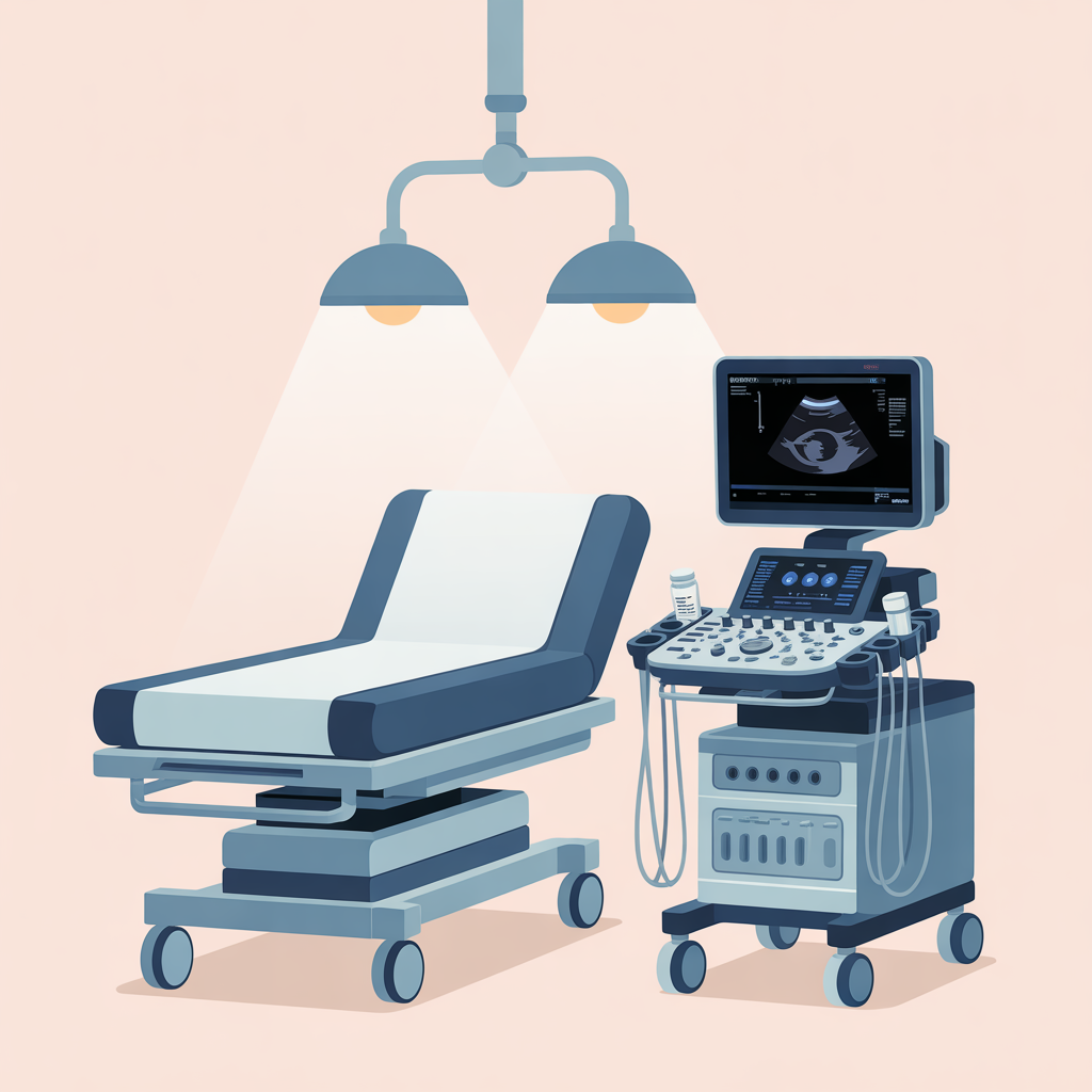
A 6-week ultrasound is typically one of the first imaging studies performed during pregnancy, occurring around 6 weeks after your last menstrual period (LMP). At this stage, healthcare providers use ultrasound technology to confirm pregnancy, assess fetal development, and rule out potential complications.
There are two primary types of ultrasound that may be used at 6 weeks:
Transvaginal Ultrasound: This is the most common method at 6 weeks because it provides clearer, more detailed images of early pregnancy structures. A small probe is gently inserted into the vagina, allowing for better visualization of the uterus and developing embryo.
Abdominal Ultrasound: Less commonly used at 6 weeks due to limited visibility, this external method involves placing a transducer on your abdomen. It may be attempted first, but transvaginal ultrasound often provides better results at this early stage.
During your appointment, you can expect the procedure to take 15-30 minutes. The technician or healthcare provider will explain what they’re seeing and may print images for you to take home.
Fetal Development at 6 Weeks
Understanding what’s happening developmentally at 6 weeks helps set realistic expectations for your ultrasound. At this stage, your baby has transformed from a cluster of cells into a recognizable embryonic structure.
Size and Appearance
At 6 weeks, your embryo measures approximately 4-6 millimeters in length, roughly the size of a sweet pea. While tiny, this represents significant growth from just a few weeks earlier. The embryo appears as a small, curved structure with a distinct head and tail end, though specific features may not yet be clearly distinguishable.
Key Developmental Milestones
Several critical developments occur around the 6-week mark:
- Neural Tube Formation: The foundation of your baby’s brain and spinal cord is developing rapidly
- Cardiovascular System: The heart begins beating, though it may not be detectable on ultrasound until slightly later
- Limb Bud Development: Early formations that will become arms and legs start to appear
- Facial Feature Formation: Basic structures for eyes, nose, and mouth begin developing
- Organ Development: Primitive versions of major organs start forming
Gestational Sac and Yolk Sac Visibility
Two important structures that should be visible on a 6-week ultrasound are the gestational sac and yolk sac. The gestational sac is the fluid-filled structure that surrounds the embryo, while the yolk sac provides early nutrition for the developing baby before the placenta takes over this function.
What You Can See on a 6-Week Ultrasound
A successful 6-week ultrasound should reveal several key structures that confirm a healthy, progressing pregnancy.
Gestational Sac
The gestational sac appears as a dark, round or oval structure within the uterus. At 6 weeks, it typically measures 18-24 millimeters in diameter. The sac should be located within the upper portion of the uterus (fundus) and have a smooth, regular border. Its size correlates with gestational age, making it useful for dating the pregnancy.
Yolk Sac
The yolk sac appears as a small, circular structure within the gestational sac. It typically measures 3-5 millimeters at 6 weeks and serves crucial functions including early blood cell formation and nutrient provision. A visible yolk sac confirms that pregnancy structures are developing appropriately.
Fetal Pole
The fetal pole is the earliest visible form of the embryo itself. At 6 weeks, it appears as a small, elongated structure adjacent to the yolk sac. The presence of a fetal pole with appropriate measurements for gestational age is a positive indicator of fetal development.
Embryonic Structures
While still very early in development, you may begin to see differentiation between the head and body regions of the embryo. The head end typically appears slightly larger, and early organ development may be detectable, though specific organs aren’t yet clearly distinguishable.
Heartbeat Detection at 6 Weeks
One of the most anticipated moments during a 6-week ultrasound is detecting the fetal heartbeat, though it’s important to understand that timing can vary.
When Heartbeat Becomes Detectable
Fetal heart activity typically becomes detectable between 5.5 and 6.5 weeks of pregnancy. However, several factors can influence detection timing:
- Exact gestational age
- Position of the embryo
- Quality of ultrasound equipment
- Maternal factors (such as weight or uterine position)
Normal Heart Rate Ranges
When detectable at 6 weeks, the fetal heart rate typically ranges between 90-110 beats per minute (BPM). This is slower than the heart rate you’ll see later in pregnancy, which typically reaches 120-180 BPM. The heart rate generally increases as the pregnancy progresses through the first trimester.
What if No Heartbeat is Detected
If no heartbeat is detected at 6 weeks, it doesn’t necessarily indicate a problem. Several scenarios may explain this:
- Pregnancy may be earlier than estimated
- Technical limitations of the ultrasound
- Natural variation in development timing
Your healthcare provider will typically recommend follow-up ultrasounds in 1-2 weeks to monitor progress before drawing any conclusions.

Measurements and Dating
Accurate measurements during your 6-week ultrasound help confirm gestational age and assess appropriate fetal growth.
Crown-Rump Length (CRL)
Crown-rump length measures the embryo from the top of the head (crown) to the bottom of the torso (rump). At 6 weeks, this typically measures 4-6 millimeters. CRL is considered one of the most accurate methods for dating early pregnancy, with accuracy within 3-5 days.
Gestational Age Confirmation
Healthcare providers compare ultrasound measurements with your estimated gestational age based on your last menstrual period. If there’s a significant discrepancy (more than 5-7 days), your due date may be adjusted based on ultrasound findings, as these measurements are typically more accurate than LMP dating.
Growth Assessment
Appropriate growth for gestational age is assessed by comparing measurements to established norms. Your healthcare provider will evaluate whether structures are measuring appropriately for the estimated gestational age and growing at expected rates.
Different Types of Results and What They Mean
Understanding potential ultrasound findings helps prepare you for various scenarios and their implications.
Normal Findings
A normal 6-week ultrasound typically shows:
- Single intrauterine gestational sac in appropriate location
- Visible yolk sac with normal size and appearance
- Fetal pole with measurements appropriate for gestational age
- Detectable fetal heart activity (though this may occur slightly later)
- No evidence of bleeding or fluid collections
Concerning Findings
Some findings may require additional monitoring or intervention:
- Absence of fetal pole when expected based on gestational age
- Irregular or unusually small gestational sac
- Absence of yolk sac when gestational sac is large enough
- Evidence of bleeding (such as subchorionic hematoma)
- Measurements significantly smaller than expected for dates
Multiple Pregnancies
Twin pregnancies can often be detected at 6 weeks, appearing as separate gestational sacs or a single sac with two yolk sacs or fetal poles. Twin pregnancies require specialized monitoring throughout pregnancy, and your healthcare provider will discuss implications and additional care requirements.
Potential Issues and Complications
While most 6-week ultrasounds reveal healthy, progressing pregnancies, it’s important to understand potential complications.
Miscarriage Signs
Early pregnancy loss affects approximately 10-20% of recognized pregnancies. Ultrasound findings that may suggest miscarriage include:
- Absence of expected structures for gestational age
- Lack of fetal heart activity when it should be detectable
- Irregular gestational sac shape or size
- Evidence of bleeding within the uterus
Your healthcare provider will typically recommend follow-up ultrasounds before confirming pregnancy loss, as dating uncertainties can affect interpretation.
Ectopic Pregnancy
Ectopic pregnancy occurs when the embryo implants outside the uterus, most commonly in the fallopian tube. Ultrasound may show an empty uterus with a gestational sac located elsewhere, though ectopic pregnancies aren’t always visible on ultrasound. This condition requires immediate medical attention as it can be life-threatening.
Blighted Ovum
A blighted ovum occurs when a gestational sac develops but no embryo forms inside. On ultrasound, this appears as an empty gestational sac without a yolk sac or fetal pole when these structures should be visible. Follow-up ultrasounds are typically recommended to confirm this diagnosis.
Preparing for Your 6-Week Ultrasound
Proper preparation helps ensure the best possible results from your ultrasound examination.
Before the Appointment
For transvaginal ultrasounds, you typically don’t need a full bladder. However, for abdominal ultrasounds, you may be asked to drink water beforehand to fill your bladder, which helps improve image quality. Bring your insurance cards, a list of current supplements or vitamins, and any previous ultrasound images or medical records.
During the Procedure
The ultrasound procedure involves:
- Checking in and completing any necessary paperwork
- Changing into a hospital gown if required
- Positioning on the examination table
- Insertion of the transvaginal probe (if applicable) or application of gel for abdominal ultrasound
- Image capture and measurements
- Discussion of findings with your healthcare provider

Questions to Ask Your Healthcare Provider
Consider asking these important questions during your appointment:
- How do my measurements compare to expected values for my gestational age?
- When should I expect to see fetal heart activity if not detected today?
- When is my next ultrasound scheduled?
- What symptoms should prompt me to contact your office?
- Are there any activity restrictions I should follow?
- When should I start or continue prenatal vitamins?
What Happens After Your 6-Week Ultrasound
Following your ultrasound, your healthcare team will provide guidance on next steps and ongoing prenatal care.
Normal Next Steps
With normal ultrasound results, you can expect:
- Scheduling your next prenatal appointment (typically around 8-10 weeks)
- Discussion of prenatal vitamin requirements
- Review of early pregnancy symptoms and management
- Information about genetic screening options
- Scheduling routine prenatal labs if not already completed
Lifestyle Recommendations
Your healthcare provider will likely recommend:
- Taking prenatal vitamins containing folic acid
- Maintaining a healthy, balanced diet
- Staying hydrated
- Getting adequate rest
- Avoiding alcohol, smoking, and certain medications
- Moderate exercise as tolerated
- Managing stress through relaxation techniques
Warning Signs to Watch For
Contact your healthcare provider immediately if you experience:
- Heavy bleeding (soaking more than one pad per hour)
- Severe abdominal or pelvic pain
- Dizziness or fainting
- Persistent nausea and vomiting preventing fluid intake
- Fever above 100.4°F (38°C)
- Sudden cessation of pregnancy symptoms
Frequently Asked Questions
Can you always see a heartbeat at 6 weeks?
No, fetal heart activity isn’t always detectable at exactly 6 weeks. Detection timing varies based on exact gestational age, equipment quality, and individual factors. If not seen at 6 weeks, follow-up ultrasounds are typically scheduled.
Is it normal to have cramping after the ultrasound?
Mild cramping after a transvaginal ultrasound is normal and usually resolves quickly. However, severe or persistent pain should be reported to your healthcare provider.
What if my dates don’t match the ultrasound?
Discrepancies between LMP dating and ultrasound measurements are common. Ultrasound dating is generally more accurate, especially early in pregnancy, and your due date may be adjusted accordingly.
Can you determine gender at 6 weeks?
No, gender cannot be determined at 6 weeks. External genital development doesn’t begin until around 7-8 weeks, and reliable gender determination typically occurs around 15-20 weeks.
How accurate is dating at 6 weeks?
Dating accuracy at 6 weeks is typically within 3-5 days when based on crown-rump length measurements, making it one of the most accurate times for pregnancy dating.
What if the baby measures smaller than expected?
Smaller measurements may indicate earlier gestational age than calculated, natural variation in growth, or potential concerns requiring monitoring. Your healthcare provider will recommend appropriate follow-up based on the degree of discrepancy.
Conclusion
A 6-week ultrasound represents a significant milestone in your pregnancy journey, offering the first glimpse of your developing baby and confirmation of a progressing pregnancy. While this examination can detect important developmental markers like the gestational sac, yolk sac, fetal pole, and often the first heartbeat, it’s important to remember that timing can vary among individuals.
Understanding what to expect during this appointment, how to interpret different findings, and when to seek additional care empowers you to actively participate in your prenatal care. Remember that every pregnancy is unique, and variations in development timing are normal.
The most important takeaway is maintaining open communication with your healthcare provider, following recommended follow-up appointments, and seeking prompt medical attention for any concerning symptoms. Your 6-week ultrasound is just the beginning of monitoring your baby’s growth and development throughout your pregnancy journey.
If you have any questions or concerns following your ultrasound, don’t hesitate to contact your healthcare provider. They are your best resource for personalized medical advice and can address any specific concerns about your pregnancy and ultrasound results.